Home

Industrial Pescăruş de mare bec desmosomes electron microscopy Aoleu Substanţial tabără

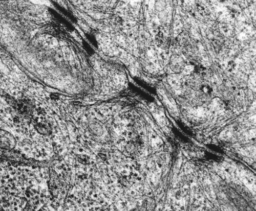
Desmosome Between Heart Muscle Cells #2 by Dennis Kunkel Microscopy/science  Photo Library
Desmosome Between Heart Muscle Cells #2 by Dennis Kunkel Microscopy/science Photo Library

About Us
About Us

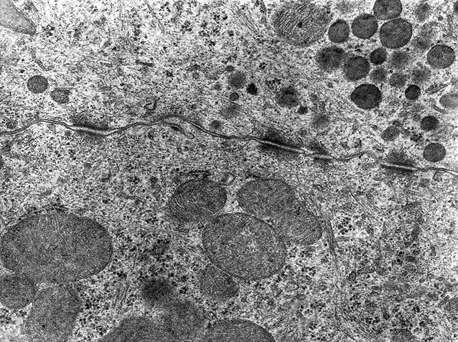
Desmosome junction, TEM - Stock Image - C049/2130 - Science Photo Library

Transmission electron microscopy of a cross section through... | Download  Scientific Diagram
Transmission electron microscopy of a cross section through... | Download Scientific Diagram

Spot desmosome Dr.Jastrow's electron microscopic atlas

Desmosome - an overview | ScienceDirect Topics

Desmosomes Tem by Alvin Telser

Chapter 18 Visualization of Desmosomes in the Electron Microscope -  ScienceDirect
Chapter 18 Visualization of Desmosomes in the Electron Microscope - ScienceDirect

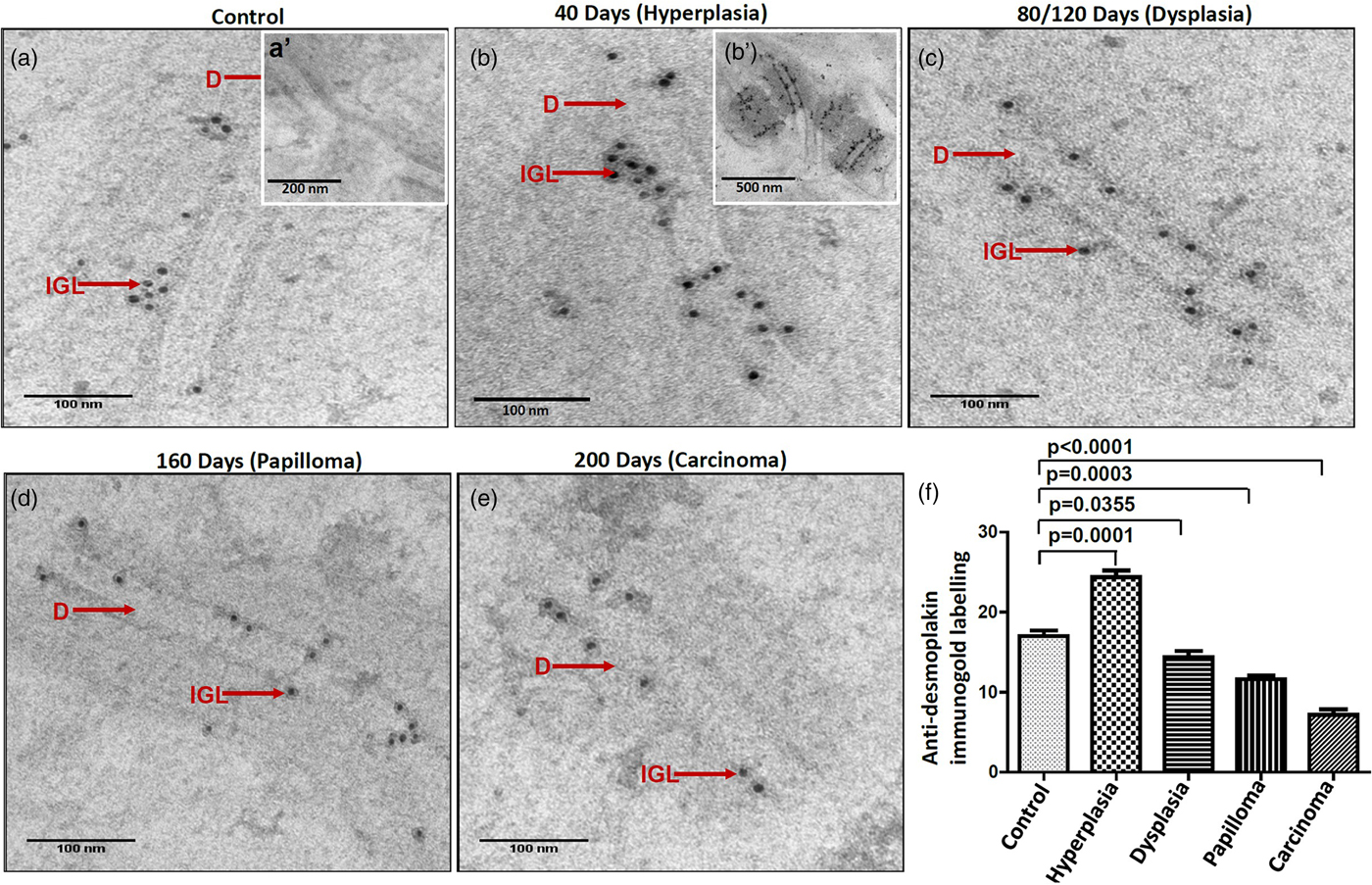
Role of Electron Microscopy in Early Detection of Altered Epithelium During  Experimental Oral Carcinogenesis | Microscopy and Microanalysis | Cambridge  Core
Role of Electron Microscopy in Early Detection of Altered Epithelium During Experimental Oral Carcinogenesis | Microscopy and Microanalysis | Cambridge Core

Spot desmosome Dr.Jastrow's electron microscopic atlas

Spot desmosome Dr.Jastrow's electron microscopic atlas

Bildagentur | mauritius images | Transmission Electron Micrograph (TEM) of  desmosomes between epithelial cells in the human cervix. Magnification:  95,000x @ 8 x 10 inches.
Bildagentur | mauritius images | Transmission Electron Micrograph (TEM) of desmosomes between epithelial cells in the human cervix. Magnification: 95,000x @ 8 x 10 inches.

basement membrane Dr.Jastrow's electron microscopic atlas

TEM of a desmosome in human epithelium - Stock Image - M132/0281 - Science  Photo Library
TEM of a desmosome in human epithelium - Stock Image - M132/0281 - Science Photo Library

Desmosomes by Jose Calvo / Science Photo Library

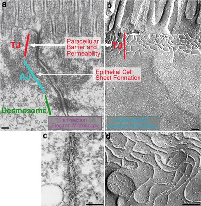
Tight junction-based epithelial microenvironment and cell proliferation |  Oncogene
Tight junction-based epithelial microenvironment and cell proliferation | Oncogene

Transmission electron micrograph illustrating desmosomes (DES) and... |  Download Scientific Diagram
Transmission electron micrograph illustrating desmosomes (DES) and... | Download Scientific Diagram

Desmosome, TEM | Stock Image - Science Source Images

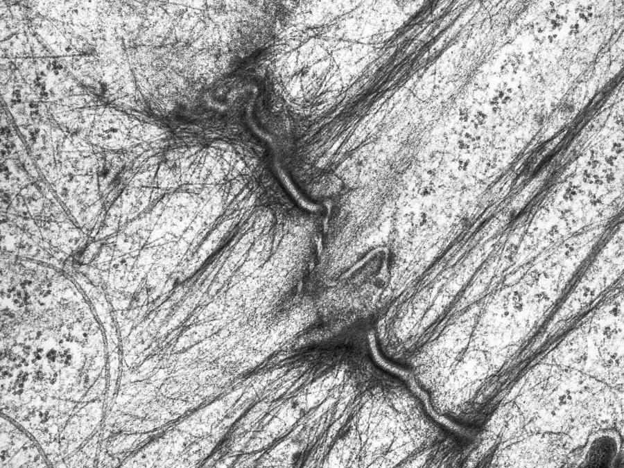
Transmission electron microscope (TEM) micrograph showing three desmosomes  (maculae adherentes) with prominent dense plaques where keratin intermediat  Stock Photo - Alamy
Transmission electron microscope (TEM) micrograph showing three desmosomes (maculae adherentes) with prominent dense plaques where keratin intermediat Stock Photo - Alamy

Desmosomal-mitochondrial associations | thank you science

Desmosomes, Desmocollin, Desmoglein, and Desmoplakin (Molecular Biology)

Desmosomes joining an intermediate cell (INT) and a basal cell (BAS) in...  | Download Scientific Diagram
Desmosomes joining an intermediate cell (INT) and a basal cell (BAS) in... | Download Scientific Diagram

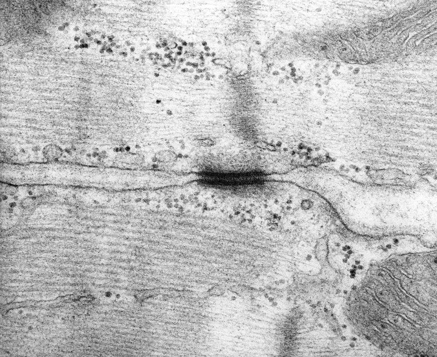
Electron microscopy of desmosome formation after 120 min-TPA (10 ng per...  | Download Scientific Diagram
Electron microscopy of desmosome formation after 120 min-TPA (10 ng per... | Download Scientific Diagram